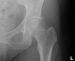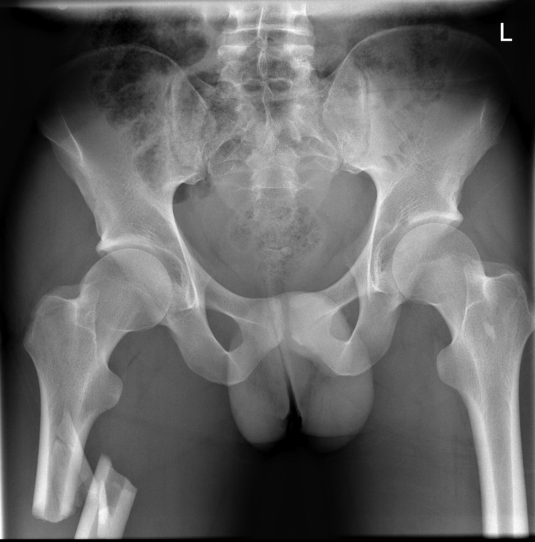
 Fracture line on the inferior surface indicate compression fracture. Fracture line is present on the superior aspect of the femoral neck on the tensile surface. The routine minimal evaluation for neck of femur fracture must include two views - anteroposterior view of the hip and pelvis and a cross-table lateral view. Radiographic imaging is important in diagnosis, classification, treatment and follow-up assessment of neck of femur fracture. X Ray X ray of Pelvis with both Hips showing right side fracture neck of femur. If unrecognized or untreated, these fractures at the femoral neck can lead to two serious. The routine minimal evaluation for neck of femur fracture must include two views - an anteroposterior (AP) view and lateral view. Radiology, Femur fracture, Femur stress, Imaging review. Radiographic imaging is important in diagnosis, classification, treatment and follow-up assessment of neck of femur fracture. Risk calculators and risk factors for Neck of femur fracture x rayĮditor-In-Chief: C. Natural History, Complications and PrognosisĪmerican Roentgen Ray Society Images of Neck of femur fracture x rayĪll Images X-rays Echo & Ultrasound CT Images MRIĭirections to Hospitals Treating Oral cancer A displaced femoral neck fracture may disrupt the blood supply to the femoral head resulting in avascular necrosis.Differentiating Neck of femur fracture from other Diseases Blood supply to the femoral head is retrograde and dependent on the femoral neck. The hip capsule inserts just proximal to the intertrochanteric line. The Garden classification of femoral neck fractures (FNF) dictates treatment via internal fixation or hip replacement, including hemiarthroplasty or total hip arthroplasty.
missed or maltreated intracapsular fractures risk avascular necrosis.dynamic hip screw if hemiarthroplasty not required.hemiarthroplasty for displaced intracapsular fractures.CT or MRI can be used to find occult minimally displaced fractures.The term 'hip fracture' is applied to fractures in any of these locations. normal radiograph with persistent symptoms needs further Ix The femoral neck connects the femoral head to the proximal portion of the femoral shaft and attaches to the intertrochanteric region ( figure 1 ).subtrochanteric: fractures below the trochanters.

trochanteric: fractures that span the intertrochanteric line Radiology Masterclass Trauma X-ray- Tutorial - Lower limb X-rays - X-rays of Hip fractures and the femoral neck, also known as neck of femur fractures or.basicervical: at the bottom ( base) of the neck ( cervical).subcapital: just below the femoral head ( capitis).femoral neck: extra-articular, intra-capsular fractures.femoral head: fractures of the head that extend to the joint.capsular insertion medial to the intertrochanteric line.division into intracapsular and extracapsular fractures.painful hip and inability to weight-bear.smaller peak in younger patients involved in high-energy trauma.high incidence of low-energy osteoporotic fractures in the elderly.incidence of 1/1000 in Western populations Femoral head fracture dislocation- Pipkin classification Subcapital femoral neck fracture- Garden classification Intertrochanteric fracture- integrity of the.For more information, you can read a more in-depth reference articles: proximal femoral fractures, femoral neck fractures.


 0 kommentar(er)
